Figure 1.
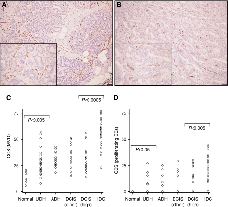
Endothelial staining in breast lesions. Immunohistochemical staining of ECs with (A) CD31 and (B) endoglin in high-grade DCIS. Note the increased frequency and intensity of stained ECs for CD31 compared with endoglin (scale=50 μm). Scatter plots representing cumulative Chalkley scores in different breast tissue samples immunostained with (C) CD31 (MVD) and (D) endoglin (proliferating ECs). (C) There was a significant increase in MVD between normal breast tissue and UDH (P<0.005), and a further significant increase in MVD between in situ (DCIS) and invasive breast cancers (P<0.0005). (D) There was a significant increase in proliferating ECs in UDH cases compared with normal breast (P<0.05) and in IDC compared with high-grade DCIS specimens (P<0.005). P<0.05 was considered significant (Kruskal–Wallis followed by Mann–Whitney U-test).

