Figure 2.
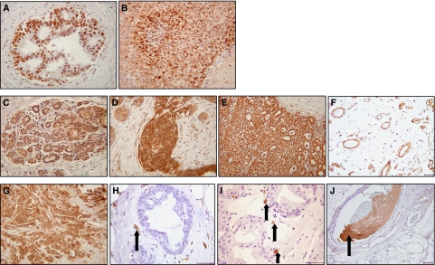
Immunohistochemical staining for HIF-1α, VEGF and TF. (A) Nuclear HIF-1α staining in the ductal epithelial cells of an ADH case and (B) tumour cells of an invasive cancer. (C) Weak expression of VEGF in normal breast epithelium (score=1), (D) strong expression in florid usual ductal hyperplasia (score=2/3) and (E) strong staining localised to tumour cells within invasive breast carcinomas (score=3). (F) VEGF expression in ECs in normal breast tissue. (G) Tumour cells expressed TF in approximately 55% of invasive breast cancer specimens. (H) TF was expressed in ECs associated with benign hyperplastic tissue (arrow). (I) Putative macrophages expressing TF associated with areas of DCIS (arrows). (J) TF expressed in vessel containing thrombosis (arrow). Photographs A–E and G were taken at × 20 magnification and all others at × 40 magnification.

