Fig. 1.
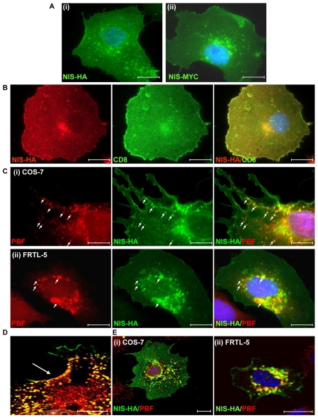
NIS and PBF localisation in COS-7 and FRTL-5 cells. (A) Following transfection into COS-7 cells and detection by immunofluorescence analysis, NIS-HA (i) and NIS-MYC (ii) demonstrated a similar pattern of expression. Both proteins were located predominantly within the plasma membrane, with partial staining within intracellular vesicles. (B) Coexpression of NIS-HA (red) with the plasma membrane marker CD8 (green) resulted in a high degree of colocalisation as seen in the merged image (yellow). (C) Representative fluorescence immunocytochemistry experiments examining staining of PBF (red) and NIS-HA (green) following transient transfection in COS-7 (i) and FRTL-5 (ii) cells. PBF is predominantly expressed within cytoplasmic vesicles. Merged images demonstrate strong colocalisation between PBF and NIS (yellow) (arrows). (D) Enlarged merged confocal image of PBF (red) and NIS-HA (green) colocalisation (yellow) at the cell surface (arrowed) of a COS-7 cell. (E) Confocal microscopy of NIS and PBF localisation in representative COS-7 and FRTL-5 cells. Colocalisation of PBF and NIS was confirmed by yellow staining in the merged images. Nuclei are visualised in blue with Hoechst 33342 stain in the merged images. Scale bars: 20 μM.

