Fig. 3.
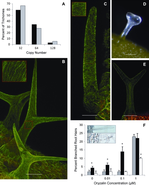
ETR2 affects microtubule assembly. (A) Both wild-type (black bar) and etr2-3 (grey bar) nuclei show the same level of endoreduplication (P <0.05, t test). (B) Wild-type trichomes expressing 35S:MAP4-GFP show a longitudinal fine mesh of microtubules (scale bar=50 μm). Inset is a magnification of longitudinally oriented microtubules in the stalk of the trichome. (C) Some etr2-3×35S:MAP4-GFP trichomes nearing maturity show disorganized microtubule cytoskeleton and early signs of depolymerization at the base of the trichome (scale bar=30 μm). Inset is a magnification of disorganized microtubule assembly discussed in the text. (D) Branching is induced in etr2-3 when treated with 20 μM paclitaxol for 2 h and then grown on MS medium until new leaves are produced (scale bar=100 μm). (E) etr2-3×35S:MAP4-GFP lines which express the highest level of MAP4-GFP show trichome branching and normal microtubule assembly (scale bar=70 μm). (F) Oryzalin causes depolymerization of microtubules and induces unbranched root hairs to become branched (inset). Bar chart of dose-response of the wild type (grey bars), etr2-3 (black bars), and etr2-1 (white bars) to different oryzalin concentrations. Asterisk indicates statistically significant difference from the wild type (P <0.05, t test).

