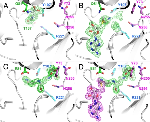
Fig. 3.
Unbiased difference Fourier maps of wild-type and variant MtNAS co-crystallized or soaked with substrate and/or products. All of the maps are contoured at 2.5σ. Residues from MtNAS lining the reaction chamber are color-coded as for Fig. 2B. (A) E81Q-MtNAS co-crystallized in the presence of 5 mM glutamate. The glutamate substrate is in the donor site and adopts two alternative positions. (B) E81Q-MtNAS co-crystallized with 5 mM glutamate and soaked in a solution containing 5 mM SAM. The aminopropyl moiety of the SAM takes the position previously occupied by the glutamate substrate. The glutamate position is however disordered and contributes to the bulky electron density in the reaction chamber. (C) Electron density map of native MtNAS co-purified with tNA. (D) Co-crystallization of native MtNAS with 5 mM MTA (the electron density associated with MTA is colored in magenta).

