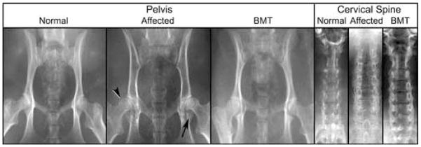
Figure 1. Composite Radiograph of MPS I Normal, Affected, and BMT-treated cats.
Images of the pelvis show characteristic changes in MPS I affected cats: coxofemoral joint subluxation (arrowhead), and acetabular flattening with bony proliferation (arrow). These changes are reduced in the BMT treated animal. The untreated MPS I cervical spine shows increased vertebral width and bony proliferation, both of which are reduced in the BMT treated animal.

