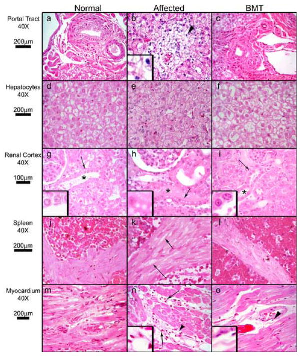
Figure 2. Histopathology of Somatic Tissues.
Hematoxylin and eosin-stained sections from normal, MPS I, and MPS I BMT-treated cats. Tissue, magnification, and micron bars located to the left of designated rows. Panels a, b, and c (liver portal tract): arrowhead (b) indicates vacuolated mononuclear cell infiltration of portal tract (3X inset) in an untreated MPS I cat, a finding absent in normal and BMT-treated cats. Panels d, e, and f (hepatocytes): the affected cat has enlarged hepatocytes with many small uniform cytoplasmic vacuoles. Normal and BMT samples have vacuolation typical of normal feline liver, are similar to each other, and differ strikingly from affected. Panels g, h, and i (renal cortex): asterisks indicate distal convoluted tubules. Cytoplasmic vacuoles in affected distal convoluted tubules are absent in normal and BMT cats (arrows indicate 3X insets). Panels j, k, and l (splenic trabeculae). Vacuolation of smooth muscle cells in MPS I (arrows) is decreased in BMT-treated cats. Panels m, n, and o (myocardium): arrows indicate perivascular infiltration by mononuclear cells in an affected cat, which is absent in the BMT animal. Arrowheads indicate 3X insets in panels n and o.

