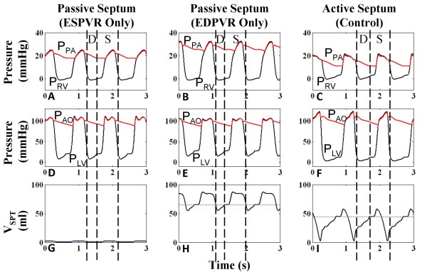
Figure 9.
Comparison of Septal Models. Ventricular and arterial pressures and septal volume for three septal models – case a: passive septum with linear end-systolic pressure-volume relationship (ESPVR) held throughout cardiac cycle (Panels A, D, G); case b: passive septum with nonlinear end-diastolic pressure-volume relationship (EDPVR) held throughout cardiac cycle (Panels B, E, H); case c: active septum with linear ESPVR and nonlinear EDPVR modulated by a septal activation function in the cardiac cycle (Panels C, F, I) (see text for details). For case a, the septum is highly noncompliant and nearly fixed at neutral position. This curtails systolic PLV and creates an abnormal upward slope in systolic PRV. Case b severely bows the septum rightward and the relatively stiff septum, whose movement is subject only to left-to-right trans-septal gradient, mirrors PLV. Systolic PRV is high due to rightward septal position during systole. With the active septum of case c, systolic ventricular pressures have opposing slopes as seen in clinical data [11]. The septum is activated at systole to produce a strong leftward thrust (D = Diastole, S = Systole).

