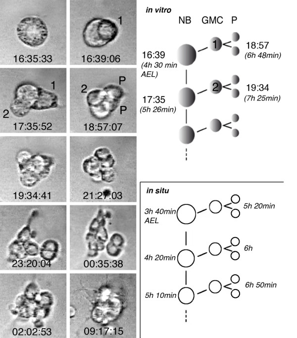
Figure 3.

Division pattern of an individually cultured neuroectodermal progenitor cell. Left panel: selected frames from time-lapse recordings (real time is indicated) of a developing neuroectodermal progenitor cell (see Additional file 4 for the corresponding movie). Isolated neuroectodermal cells show a stem cell mode of division, which is typical for neuroblasts (NB). With each of their asymmetric divisions they self renew and bud off a smaller daughter cell (ganglion mother cell (GMC); the numbers 1 and 2, indicate the first two GMCs), which typically divides one more time into two postmitotic progeny (P). The size of the NB is approximately 10 μm and of postmitotic progeny cells 5 to 6 μm. Right panel: the time points when the first divisions took place in this particular lineage (grown at 22°C) are indicated at the top (compare left panel). The respective times after egg laying (AEL) are indicated in brackets. Note that the cell cycle of GMCs is significantly longer than that of the NB. The lower scheme shows for comparison the timing (at 25°C) of the first mitotic cycles of NBs and GMCs as observed in situ (according to Hartenstein et al. [69]).
