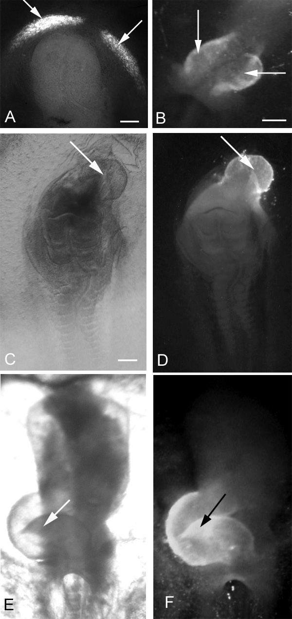
Fig. 1.

Effects of exogenous HCy on early chick heart development after a 24-hour incubation. Immunolocalization of MF20 defines the presence of cardiac tissue (arrows). (A,B) Two extremes of cardiabifida: in A, two small cardiogenic regions are differentiating bilaterally; in B, the two heart fields have moved close to the midline, are almost touching, and are ready to fuse. (C) Light microscopic view of an embryo displaying a severely truncated neural tube with cardiac tissue differentiating cephalad to the neural area (D). (E) Bright-field ventral view of a normal, control, right-looping heart and (F) fluorescence view showing MF20 localization in the heart shown in E. In all panels, the embryonic anterior is at the top. Bars, 300 μm.
