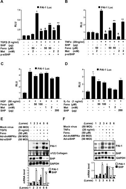
Figure 6. Inhibition of cytokine-induced PAI-1 promoter activity by fenofibrate is SHP-dependent.
A-D: HepG2 cells were transfected with the human PAI-1 gene promoter (−800bp) for 18 h followed by treatments with TGFβ (panel A), TNFα (panel B), HGF (panel C) and IL-1α (panel D) at indicated concentrations for 24 h in the presence or absence of SHP (200 μg), pSuper siRNA SHP (p-siSHP, 200 μg), fenofibrate and metformin for another 24 h under serum-starved conditions. All experiments were done in triplicate, and data are expressed in relative luciferase units (RLU) and as the fold activation relative to the control, representing mean ± SD of 3 individual experiments.*P < 0.05, &P < 0.05 and **P < 0.005, compared to untreated control, individual cytokine treatments and fenofibrate or metformin treated cells. E-F: HepG2 cells were treated with mock virus, adenovirus siRNA SHP (Ad-siSHP) and adenovirus dominant negative AMPKα (Ad-dnAMPKα) for 48 h, followed by fenofibrate (Feno) treatment at indicated concentration for an additional 24 h, after which cells were treated with TGFβ (panel E), TNFα (panel F), at indicated concentrations for another 4 h under serum starved conditions. Total RNA was isolated for Northern blot analysis of PAI-1, Collagen type I (Col I) and SHP mRNA expression and was normalized to GAPDH expression. Data represent mean ± SD of 3 individual experiments. *P < 0.05, &P < 0.005 and **P < 0.005 compared to untreated control, cytokine treated cells and fenofibrate treated cells.

