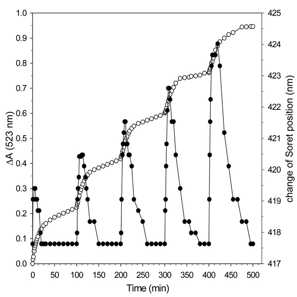
Figure 9. Binding of Bfd to BfrB promotes heme mediation of electron transfer to the core iron.
Open circles, left axis: time dependent changes in the intensity of absorption measured at 523 nm upon addition of aliquots of NADPH containing 15% of the total electron equivalents needed to reduce all the ferric ions in the core of BfrB (600 Fe3+ ions/BfB) to a solution containing BfrB (0.375 µM), Fpr (15 µM), apo-Bfd (15 µM ) bipy (3 mM). Dark circles, right axis: time dependent changes in the position of the heme Soret band following addition of each NADPH aliquot. The measurements were conducted at 22 °C.

