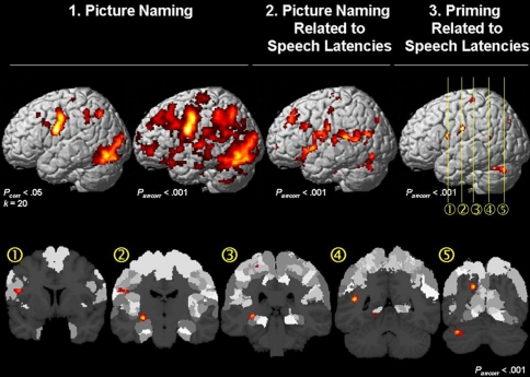
Fig. 3.
Top Left: Surface rendering of the main effect for picture naming in the fMRI data at an FWE-corrected (Pcorr < 0.05) and uncorrected (Puncorr < 0.001) threshold. Top Middle fMRI signal that is correlated with the speech latencies. Top Rightand Bottom Within the brain regions where the fMRI signal correlated with the speech latencies (Top Middle), the main effect for priming (i.e. homogeneous vs. heterogeneous trials in each condition) was significant (Puncorr < 0.001) in (1) area 44 in the left inferior frontal gyrus; (2) area 6 in the precentral gyrus; (3) the hippocampus and the postcentral gyrus (area 3b); (4) the posterior middle temporal gyrus; and (5) precuneus and the cerebellum

