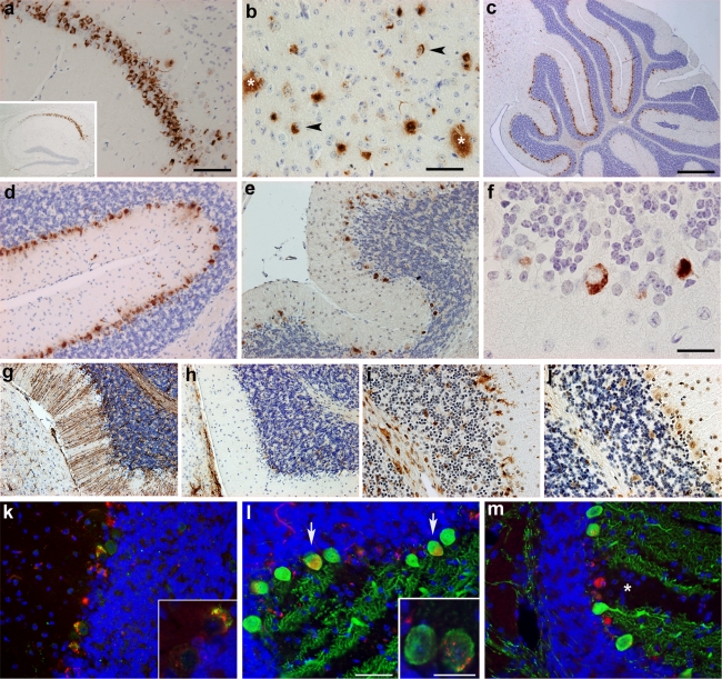
Fig. 4.
Immunohistochemical staining of TBA2 mouse brain (2-month-old). a Immunostaining with 4G8 revealed strong Aβ accumulation in the CA1 pyramidal layer of the hippocampus (inset shows a hippocampus overview at low magnification). b Intra- (arrowhead) and extracellular Aβ (asterisk) in the thalamus shown by 4G8 staining. c, d Aβ staining (4G8) in the cerebellum is almost completely restricted to the Purkinje cell layer. e, f Most Purkinje cells accumulated pyroglutamate-Aβ as shown by an antibody against Aβ3(pE). g GFAP staining of a TBA2 mouse revealed prominent Bergmann glia immunoreactivity, whereas wildtype animals (h) were consistently negative. The microglia marker Iba1 revealed microglia clusters surrounding Purkinje cells and in white matter tracts in TBA2 mice (i), but not in wildtype littermates (j). k Immunostaining of Purkinje cells with 4G8 (red) and anti-ubiquitin (green) antibodies showing abundant ubiquitin immunoreactivity in 4G8-positive Purkinje cells. l, m Staining of Purkinje cells using antibodies against calbindin (green) and 4G8 (inset shows high magnification of a 4G8- and calbindin-positive Purkinje cell). Note absent calbindin (asterisk) and extracellular Aβ staining indicating Purkinje cell loss. Only 4G8-positive remnants can be seen. Scale barsa, d, e, g–j 100 μm; b, k–m 50 μm; c 500 μm; finsetk, l 20 μm

