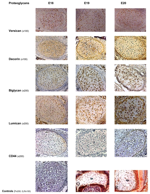
Fig. (1).
Immunoperoxidase staining for Versican showing positive staining of moderate intensity on Meckel’s cartilage chondrocytes and weak staining within the ECM at all embryonic stages (A, B, C). Decorin showed an intense positive staining on chondrocytes and within the ECM at all embryonic stages (D, E, F). Biglycan was moderately expressed at E18 (G), and more intense staining at later stages (H, I). Staining for Lumican on the chondrocytes and ECM was weak at E18 (J), moderate at E19 (K), and intense at E20 (L). Immunoperoxidase staining for CD44 showed very weak staining on chondrocytes at all stages, without any staining within the ECM (M, N, O). P, Q and R represent the control sections.
P: negative control section; Q: positive staining for biglycan in the enamel organ and dental papilla of a developing molar tooth bud at the E19 stage; R: positive staining for decorin in the dental papilla, odontoblasts and dentin in a developing tooth bud. OOE: outer enamel epithelium; SR: stellate reticulum; IEE: inner enamel epithelium; dp: dental pulp, df: dental follicle.

