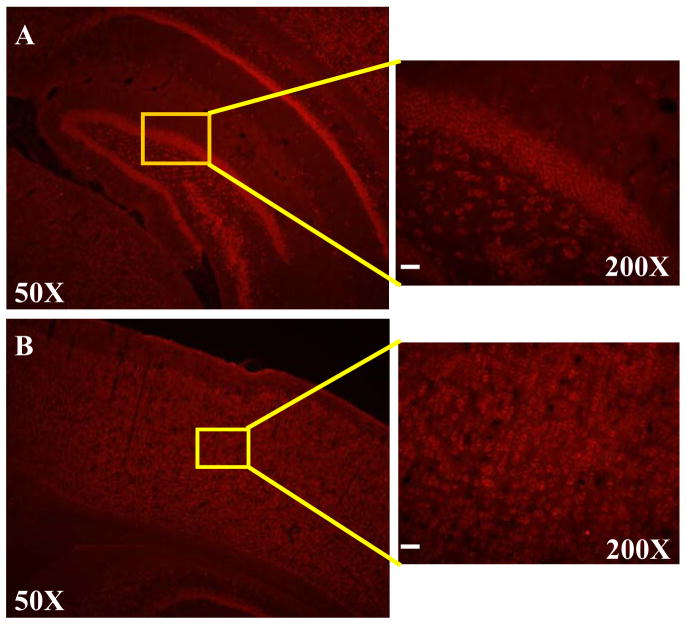
Fig. 4.
LRRK2 is highly expressed in the cortical and the hippocampal regions. Rat brain sections were incubated with anti-LRRK2 antibody. Shown in the left column are representative fluorescence photographs of LRRK2 immunohistochemistry from rat brain at the hippocampus (A) and cortex (B) levels. In the right column, high magnification images corresponding to boxed areas from the overview image are shown for rat brain, hippocampal region (A) or cortical region (B). Scale bar=40μm.

