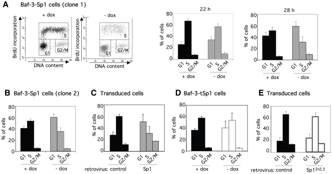
Figure 7. Cell cycle inhibition induced by Sp1 requires its binding to DNA.
(A) Baf-3-Sp1 clone 1 was grown in presence of doxycycline (+dox) or in absence of doxycycline for the indicated times (- dox) and pulsed with BrdU for 20 min. Cells were harvested and processed for BrdU labelling and DNA content staining. Cell cycle distribution was analysed by flow cytometry. Representative cell cycle profile with or without doxycycline at 28 hrs (left panel). Percentage of cells in the different phases of the cell cycle 22 or 28 hrs after treatment (right panel). (B) Cell cycle distribution of Baf-3-Sp1 clone 2 at 72 hrs. (C) Baf-3 cells were transduced with control or Sp1 encoding retrovirus. Cell cycle distribution among transduced (CD2 positive) cells 30 hrs post-infection. (D) Cell cycle distribution of Baf-3-tSp1 clone analysed after 72 hrs. (E) Baf-3 cells transduced with control or Sp1Zn2,3 encoding retroviruses were analysed 30 hrs later. Percentage of cells in the different phases of the cell cycle assessed among CD2 positive cells. Results show the means of ± sd of 2 to 3 independent experiments.

