Figure 3.
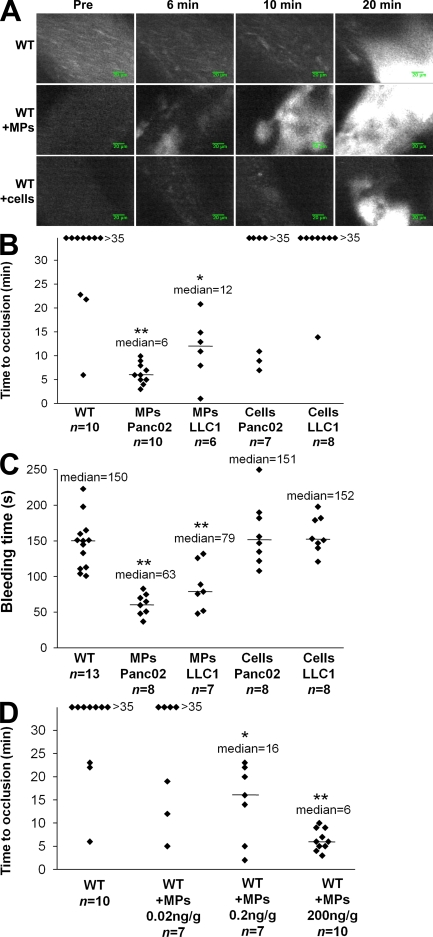
Cancer cell–derived MPs accelerate thrombus formation in vivo. Thrombus formation was assessed after infusion of Alexa Fluor 660–conjugated anti-CD41 antibody into wild-type mice in the presence of Panc02-derived MPs (MPs Panc02, 0.2 µg of MP-associated proteins/g/mouse), LLC1-derived MPs (MPs LLC1, 0.08 µg of MP-associated proteins/g/mouse), or Panc02 cells (Cells Panc02, 5,000 cells/g/mouse) or LLC1 cells (Cells LLC1, 5,000 cells/g/mouse). Injury of the mesenteric vessels was induced with 10% FeCl3 for 5 min. (A) Representative composite images of fluorescence depicting thrombus formation of labeled platelet accumulation (white) on mesenteric vessels (Pre, before injury). (B) Time to vessel occlusion reported in minutes for mesenteric arterioles in wild-type mice (n = 10 thrombi in 10 mice), wild-type mice infused with Panc02-derived MPs (n = 10 thrombi in 7 mice), or with LLC1-derived MPs (n = 6 thrombi in 6 mice), Panc02 cells (n = 7 thrombi in 7 mice), or LLC1 cells (n = 8 thrombi in 8 mice). (C). Bleeding time in seconds determined in wild-type mice (n = 13 mice), wild-type mice infused with Panc02-derived MPs (n = 8 mice), or with LLC1-derived MPs (n = 7 mice), Panc02 cells (n = 8 mice), or LLC1 cells (n = 8 mice). (D) Times to arteriole occlusion were observed in wild-type mice (n = 10 thrombi in 10 mice) and in wild-type mice infused with different concentrations of Panc02-derived MPs (MPs 0.02 ng/g/mouse, n = 7 thrombi in 6 mice; MPs 0.2 ng/g mouse, n = 7 thrombi in 7 mice; and MPs 200 ng/g mouse, n = 10 thrombi in 9 mice). Horizontal bars indicate median values. Experiments were independently performed 10 times (A and B) and at least six times (C and D). **, P < 0.01; *, P < 0.05.

