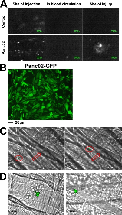
Figure 6.
Endogenous cancer cell–derived MPs accumulate at the site of thrombus formation in vivo. (A) Qdot-labeled Panc02 cells (2 × 10⁶ cells) or Qdots alone (Control) were injected subcutaneously into the right flank of a mouse. 1 wk later, fluorescent signals were detected at the site of injection (left), and in the mesentery before (middle panel) and after (right) FeCl3-induced injury. (B) Fluorescence microscopy of Panc02 cells overexpressing GFP before subcutaneous injection into wild-type mice. (C and D) 2 × 10⁵ Panc02 cells overexpressing GFP were injected subcutaneously into the right flank of a mouse. 5 wk later, the cremaster was isolated and fluorescent circulating GFP-labeled MPs were detected in the blood microcirculation (red arrow, direction of the blood flow; red circle, GFP microparticle; C) and accumulating at the site of injury in venules (D, left) and arterioles (D, right). All images are representative of three independent experiments observed for nine thrombi formed in three mice.

