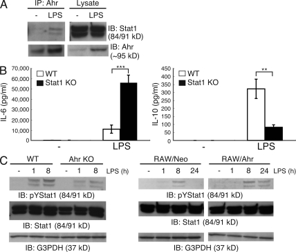
Figure 3.
Association between Ahr and Stat1 in macrophages. (A) Peritoneal macrophages were isolated from BALB/c mice and stimulated by LPS for 24 h. Interaction between Ahr and Stat1 was examined by means of immunoprecipitation (IP) and Western blotting. Data are from one representative of three independent experiments. IB, immunoblot. (B) WT and Stat1 KO peritoneal macrophages were stimulated with LPS. Supernatants were collected 24 h after stimulation, and the production of IL-6 and IL-10 were measured by means of ELISA. Data show means ± SEM of three independent experiments (**, P < 0.005; ***, P < 0.001). (C) WT and Ahr KO peritoneal macrophages or RAW/Neo and RAW/Ahr cells were incubated with LPS at the indicated time points. Whole-cell lysates were used for immunoblotting analysis with anti–phospho-tyrosine Stat1, Stat1, and G3PDH antibodies (Ab). Data are from one representative of three independent experiments.

