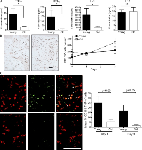
Figure 6.
Levels of TNF-α are reduced in blister fluid and in CD163+ macrophages in old skin. (A) Skin suction blisters were raised over the site of C. albicans antigen injection at day 3. Levels of TNF-α, IFN-γ, IL-10 and IL-6 were measured in the blister fluid using cytometric bead arrays. The results shown are the mean and SEM of 6 young and 10 old subjects for each cytokine tested. A significant reduction in the level of TNF-α, IFN-γ, and IL-6 was noted in the old group compared with the young group (Mann-Whitney test; *, P = 0.01 for TNF-α and IFN-γ; *, P = 0.03 for IL-6). The number of CD163+ cells within the dermis was determined by indirect immunoperoxidase staining of 6-µm tissue sections. (B, left) Representative staining of one young and one old subject 3 d after C. albicans injection. We investigated a total of five young and six old volunteers, and the collective results are shown (B, right). We also investigated the presence of CD163+ macrophages at different times after C. albicans injection (B, right). Graph shows mean number of CD163+ macrophages before injection (n = 5 young, 5 old), day 1 (n = 5 young, 7 old), and day 3 after injection (n = 5 young, 6 old). The data represent the mean ± SEM. Bar, 100 µm. Double immunofluorescence staining of TNF–α (green) and CD163 (red) in skin biopsies 3 d after C. albicans injection. Results shown are representative of one young and one old subject (C, left). These experiments were performed in three young and three old subjects and the collective results showing the mean and SEM of the percentage of CD163+ macrophages producing TNF-α per perivascular infiltrate are shown in (C, right). Bar, 100 µm.

