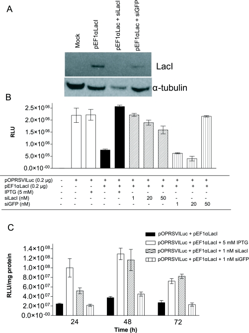
Figure 4.
Silencing of lac repressor restores luciferase expression. A. 5x10⁵ PC-3 cells were transfected with 2μg of pEF1α-LacI vector using Lipofectamine. After 2hr 100nM siLacI or siGFP complexed with Oligofectamine were added to cells. Expression of LacI was analysed 48hr after DNA delivery by western blotting using an anti-LacI antibody diluted 1/1000. Expression of α-tubulin was determined using an anti-α-tubulin antibody diluted 1/1000 as a loading control. [Key: Lane 1; mock, Lane 2; pEF1αLacI, Lane 3; pEF1αLacI + siLacI, Lane 4; pEF1αLacI + siGFP]. B. 4x10⁴ PC-3 cells were transfected with i) 0.2μg pOPRSVI-Luc alone (white bars), ii) 0.2μg pOPRSVILuc and 0.2μg pEF1α-LacI (black bars) or iii) 0.2μg pOPRSVILuc, 0.2μg pEF1α-LacI and various doses of siRNA as indicated (siLacI diagonal striped bars, siGFP vertical striped bars). Control cells received 5mM IPTG after 24hr and luciferase expression was measured after 48hr. C. 4x10⁴ PC-3 cells were co-transfected with 0.2μg pOPRSVI-Luc and 0.2μg pEF1α-LacI in the presence or absence of siRNA. Selected cells were exposed to 1nM siLacI or 1nM siGFP and luciferase activity measured at the indicated time point after siRNA delivery.

