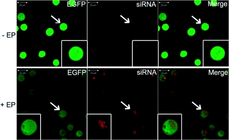
Figure 3.
Localization of anti eGFP siRNA labeled with Alexa Fluor 546 48h after electrotransfer in vitro. Cells in suspension were incubated in the presence of 1.4 μM siRNA in the pulsing buffer. Ten pulses of 5 ms at frequency of 1 Hz were applied at 0.7 kV/cm. 48 hr after siRNA electrotransfer (+EP) (lower panels), cells were sorted out by flow cytometry and were observed by confocal microscopy with ×40 objective. A zoomed picture of one cell is displayed in small boxes. Non-electrotransfected cells (-EP) were observed (top) using the same acquisition parameters. eGFP constitutively expressed in cells was detected with a 488 nm Argon laser (panels on left). Alexa Fluor 546-labeled siRNAs were detected with a 514 nm Helium-Neon laser (central panels). A merged image of the two (panels on right) points to the cytoplasmic localization of the siRNA.

