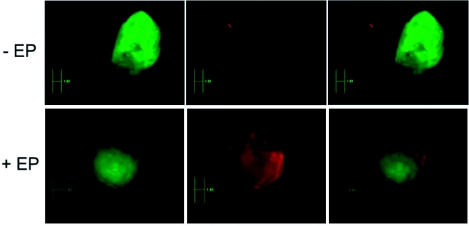
Figure 5.
Observation of the anti-eGFP siRNAs labeled with Alexa Fluor 546 after electrotransfer in tumors. 48 hr after electrotransfer (+EP) the tumors were removed and observed under stereo-microscopy, and were compared with the non-electrotransfected tumors (-EP). Left panels, constitutively expressed eGFP; middle panels, siRNA labeled with Alexa Fluor 546; right panels, merged image of left and middle panels (which emphasizes uniform distribution of the siRNA in the tumor).

