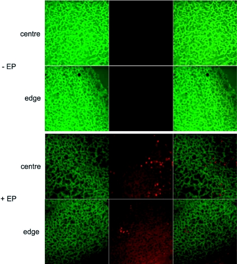
Figure 6.
Localization of the anti eGFP siRNAs labeled with Alexa Fluor 546 after electrotransfer in tumors. Forty eight hours after the electrotransfer of the siRNA (+EP) (lower half), the tumors were removed, sliced and observed under a confocal microscope with ×40 objective at 0.7 magnification. Non-electrotransfected tumors (-EP) were (top half) compared with electrotransfected tumors. The eGFP expression in cells was detected with a 488 nm Argon laser (left) and the siRNAs labeled with Alexa Fluor 546 were detected with a 514 nm Helium-Neon laser (centre). A merged image of the left and middle panels is shown on in the right panels in order to localize siRNAs.

