
Fig. 1.
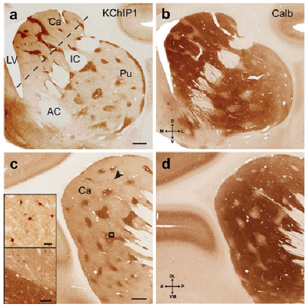
KChIP1 is a novel immunocytochemical marker for the striosomes in macaque. Coronal sections at the level of the anterior commissure showing KChIP1 immunoreactivity in the striosomes (a), with an adjacent section (b) showing immunoreactivity for calbindin in the complementary matrix compartment. In sections approximately parallel to the internal capsule (dashed line in a), a lattice-like pattern of KChIP1 immunoreactive striosomes is evident in the head of the caudate nucleus (c) with complementary staining for calbindin in an adjacent section (d). Bottom inset in c is higher magnification of box in c, and shows KChIP1immunostaining of somata in both matrix and striosomes, with heavy neuropil staining only in the striosomes. Top inset in c shows immunoreactive somata and proximal processes corresponding to the arrowhead. Scale bars: 1 mm (a and c); 40 microns (top inset in c); 100 microns (bottom inset in c). Abbreviations: AC, anterior commissure; Ca, caudate nucleus; IC, internal capsule; LV, lateral ventricle; Pu, putamen.