
Figure 1. Loss of β-catenin expression in the ventral pallidum impairs growth of the MGE.
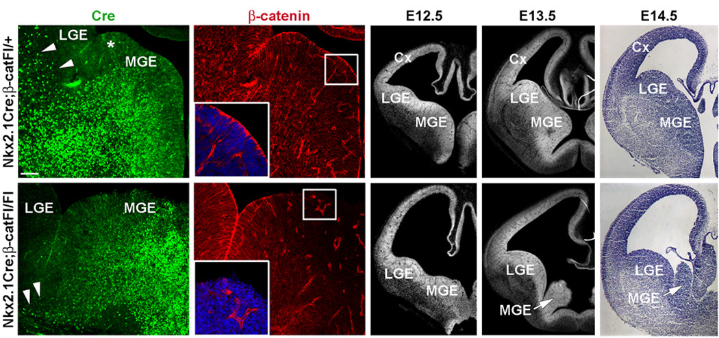
(a–d) Coronal sections from E12.5 Nkx2.1Cre;β-catFl/+ control (top row) and Nkx2.1Cre;β-catFl/Fl mutants (bottom row) co-labeled by immunofluorescence for Cre (a, c) and β-catenin (b, d). In Nkx2.1Cre mice Cre is strongly expressed through most of the MGE, with the exception of the dorsal most region (asterisk in a). Cre is also detected in cells that appear to be streaming from the MGE dorsally toward the developing striatum and cortex (arrowheads in a). This stream is greatly reduced in the mutant section (arrowheads in c). (b) Apical localization of β-catenin expression in neuroepithelial cells surrounding the lateral ventricle (insert shows higher magnification of the boxed area). (d) Consistent with the expression of Cre, β-catenin is greatly reduced in the ventral two-thirds of the mutant MGE (insert shows higher magnification of the boxed area). (e–j) DAPI (e, f, h, i) and Nissl (g, j) staining of coronal sections at E12.5, E13.5 and E14.5. After E12.5, little growth of the MGE (arrows in i and j) occurs. MGE-medial ganglionic eminence, LGE-lateral ganglionic eminence, Cx-cortex. Scale bar: 50 µm (a–d); 200 µm (e–j).