
Figure 3.
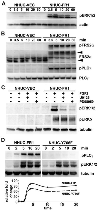
FGFR1 activates the MAPK pathway. Cells were cultured in supplement-free medium for 1 hour prior to culture with heparin and FGF2 for indicated times. A, Protein lysates were blotted with anti-pERK antibodies then reprobed with anti-actin. B, Protein lysates were blotted with phospho-specific antibodies against FRS2α and PLCγ. Blots were reprobed with anti-FRS2α or PLCγ antibodies. C, Cells were cultured with DMSO (control), U0126 or PD98059 for 1 hour. FGF2 was added for 5 minutes and lysates probed with anti-phospho ERK5 antibody that also cross-reacts with pERK1/2. Blots were reprobed with tubulin. D, NHUC-FR1 and NHUC-Y766F were cultured with FGF2 and lysates blotted for pPLCγ or pERK. Loading contol, tubulin. Graph represents the change in pERK levels in NHUC-FR1 (solid line) and NHUC-Y766F (dashed line) quantified from a representative experiment.