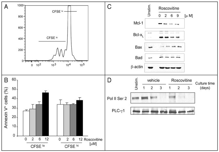
Figure 2.
Roscovitine increases apoptosis of proliferating cells by altering expression of Mcl-1 and Bax. (A and B) CFSE-labeled T cells were stimulated with anti-CD3 and anti-CD28 antibodies for 48 hrs and viability of proliferating cells (CFSElo) and non-proliferating cells (CFSEhi) was determined by expression of Annexin V. Results shown in (B) represent mean values of two independent experiments (p = 0.02). (C and D) Purified T cells were stimulated with anti-CD3 and anti-CD28 antibodies in the absence or the presence of roscovitine. Cells were cultured for 48 hours with the indicated concentrations of roscovitine (C) or with 12 μM roscovitine for the indicated time points (D), cell lysates were prepared and protein expression was analyzed by SDS-PAGE and immunoblot with the indicated antibodies. Immunoblots for b-actin and PLC-g1 were used as loading control for (C and D), respectively.

