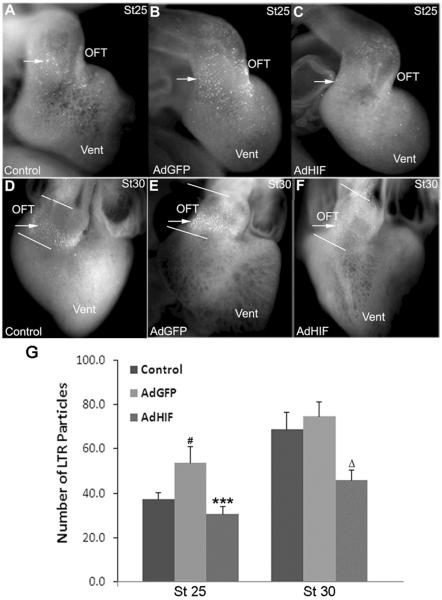
Figure 1.
Adenoviral mediated expression of HIF-1α reduces LTR staining in the cardiac OFT. LTR staining (arrows) is significantly reduced in the OFT of AdHIF-1α injected hearts (C and F) as compared to AdGFP (B and E) at both stages 25 and 30, and un-injected embryos at stage 30 (D). (G) Number of LTR particles in the OFT in whole mount as quantified by Image J. # P<0.01, compared to control; *** P<0.001 compared to AdGFP group; Δ P<0.001 compared to both control and AdGFP group. OFT, outflow tract; Vent, ventricles.

