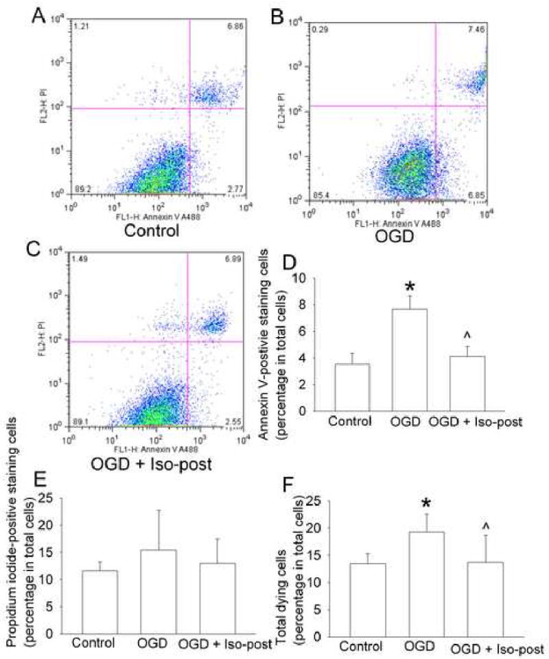
Fig. 4. Effects of isoflurane postconditioning on cell necrosis and apoptosis induced by oxygen-glucose deprivation.

Bovine pulmonary arterial endothelial cells were exposed to or not exposed to a 3-h oxygen-glucose deprivation (OGD) and posttreated with or without 2% isoflurane (Iso-post) for 1 h immediately after the oxygen-glucose deprivation. These cells were stained with annexin V and propidium iodide (PI) 3 h after the oxygen-glucose deprivation and then were analyzed by flow cytometry. A representative of sorted cells by flow cytometry for each experimental condition is presented in panel A, B and C. These are two-parameter dot plots with the x-axis showing the intensity of annexin V staining and y-axis showing the intensity of PI staining. The location of a particular cell in the plot is determined by its intensities from those two stains. The pooled results from 8 flow cytometrical studies are presented in panel D, E and F. Total dying cells are cells stained positively with annexin V and/or PI. Results are means ± S.D. * P < 0.05 compared with control. ˆ P < 0.05 compared with oxygen-glucose deprivation only.
