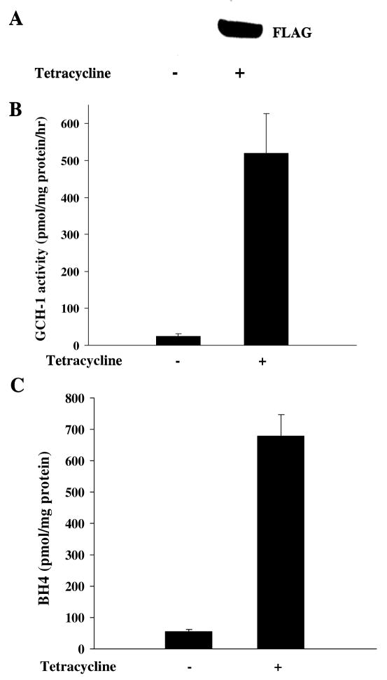
Figure 1.
Characterization of the cell line stably expressing FLAG-tagged rat GCH-1. FLAG-GCH-1 HEK 293 cells were stimulated with or without tetracycline (1μg/ml) for 24 hours. (A) Whole cell extracts were analyzed by Western blot analysis for the expression of GCH-1 using anti-FLAG antibody. (B) Cells were lysed for GCH-1 activity assay. (C) Cells were lysed and intracellular BH4 levels were measured by HPLC. Data are expressed as mean±SD (n=3). *P<0.05 vs. the cells without tetracycline treatment.

