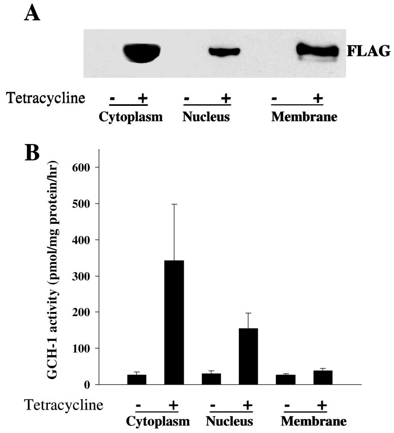
Figure 3.
Subcellular distribution of GCH-1 interacting proteins. Cell fractional proteins (cytoplasmic, nuclear and membrane proteins) from GCH-1 HEK cells were prepared using specific extraction reagents from Pierce Biotechnology. (A) 30μg proteins from each fraction were loaded on the SDS-PAGE and immunoblotted with anti-FLAG. (B) 50μl cell lysates from each fraction were analyzed for GCH-1 activity and normalized by their protein concentration. n=3/experiment

