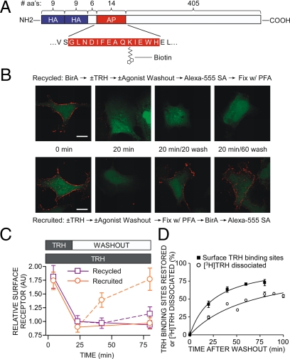
Figure 9.
Recycling of biotinylated TRH receptor. A, Model of 2HA-AP-TRH receptor. B and C, Cells expressing 2HA-AP-TRH receptor were incubated with or without 100 nm TRH with or without washout, as shown in the middle of panel B. Cells were fixed with paraformaldehyde (PFA), biotinylated with BirA, and labeled with Alexa Fluor 555 streptavidin in the order shown above (recycled) or below (recruited) the micrographs. Control experiments established that staining of naïve cells was comparable when fixation preceded incubation with BirA or when the order was reversed. Bar, 10 μm. C, Quantification by line scan of relative surface receptor. Shown are mean ± se of four to seven cells from four independent experiments. Solid lines show results of cells exposed to TRH throughout, and dashed lines show cells after TRH washout. D, To measure restoration of TRH binding sites after internalization, cells stably expressing 2HA-TRH receptors were incubated with or without 100 nm TRH for 20 min to cause receptor internalization, washed, and incubated for various times. Dishes were placed on ice, and the number of TRH-binding sites on the cell surface was titrated by incubating cells with 10 nm [3H]MeTRH on ice as described in Materials and Methods. To measure receptor cycling, cells were incubated with [3H]TRH for 20 min, washed, and incubated for different times before determination of the fraction of [3H]TRH released as described in Materials and Methods. Points show the mean ± se from five to nine experiments, each performed in duplicate. Error bars were within symbol size where not visible. aa, Amino acids.

