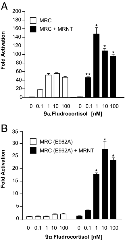
Figure 1.
9α-Fludrocortisol dose response for the MR N/C-interaction. COS-1 cells were transiently transfected with expression vectors and the GAL4-responsive luciferase reporter vector g5-LUC. The cells were treated 14–16 h after transfection with various concentrations of 9α-fludrocortisol as indicated. Luciferase activity was measured after a 24-h incubation and is represented as fold activation relative to luciferase activity in the absence of ligand. A, 9α-Fludrocortisol dose response with wild-type MRC. The open bars correspond to GAL4-MRC + pVP16, and the solid bars correspond to GAL4-MRC + pVP16-MRNT. B, 9α-Fludrocortisol dose response with MRC(E962A). The open bars correspond to GAL4-MRC(E962A) + pVP16, and the solid bars correspond to GAL4-MRC(E962A) + pVP16-MRNT. Each data point represents the mean ± sem derived from three independent experiments; *, P < 0.001; **, P < 0.01 denotes significance when compared with the same concentration of 9α-fludrocortisol in the absence of MRNT.

