
Figure 3.
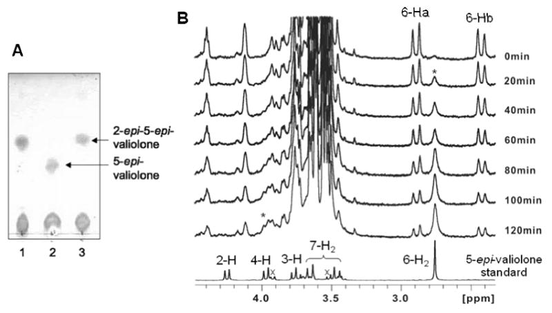
TLC and 1H NMR analyses of ValD-catalyzed reactions. A, TLC analysis of ValD-catalyzed reactions: lane 1, sedoheptulose 7-phosphate with 2-epi-5-epi-valiolone synthase ValA only; lane 2, sedoheptulose 7-phosphate with ValA and ValD; lane 3, sedoheptulose 7-phosphate with ValA and boiled ValD. B, 1H NMR analyses of ValD reaction. A solution of 2-epi-5-epi-valiolone in D2O was placed in a 5 mm NMR tube and the reaction was started by adding potassium phosphate buffer (pH 7.4, 25 mM) and purified ValD to a final concentration of 0.018 mg/ml. After one hour, additional ValD was added into the reaction mixture to a final concentration of 0.054 mg/ml protein. The geminal H-6a and H-6b of 2-epi-5-epi-valiolone appear as doublets, whereas those of 5-epi-valiolone appear as a singlet. * indicates the growing signal corresponding to the increased production of 5-epi-valiolone over the period of NMR measurement; × indicates signals of impurities.