Fig. 3.

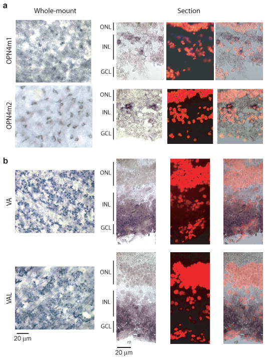
Melanopsin but perhaps not VA/VAL opsin was expressed in HC-layer of catfish retina. a, In situ hybridization of OPN4m1 and OPN4m2 mRNA. Left, digoxigenin (DIG) staining of flat-mount retina. Small, dark-blue areas surrounding hollow cores (presumably the nuclei) indicate stained cell cytoplasm. Right, Same experiments on retinal cross-sections showing DIG signal, propidium iodide (PI) nuclear staining to mark retinal layers, and the two merged. Catfish retinal layers are not very distinctive, with sparse nuclei in the inner nuclear layer7. b, In situ hybridization of VA/VAL opsin mRNA. Left, DIG staining of flat-mount retina. Dark-blue dots are the stained cells, situated close to the retina inner surface with blood vessels (arrows) evident in the same focal plane. Right, Same experiments on retinal cross-sections. In both a and b, cells appear smaller in the flat-mount retina than in retinal cross-sections owing to some tissue shrinkage during processing. ONL, outer nuclear layer; INL, inner nuclear layer; GCL, ganglion cell layer.
