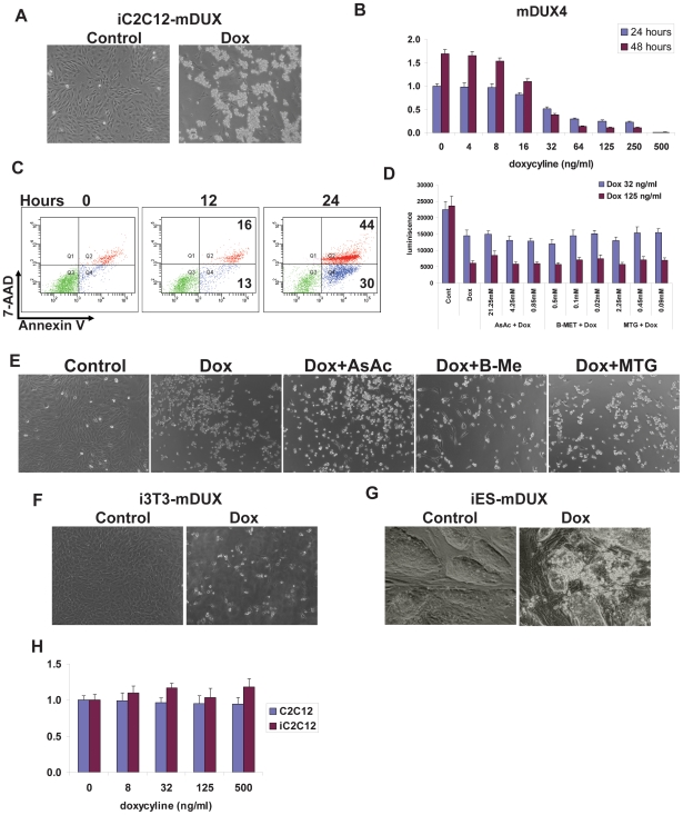
Figure 1. Toxicity of mDUX.
(A) Morphology of iC2C12-mDUX cells induced for 24 hours (Dox) with 500 ng/ml doxycycline. The majority of induced cells were detached and floating after 24 hours. (B) ATP assay for analysis of viability in iC2C12-mDUX cells induced with various concentrations of doxycyline for 24 and 48 hours. Decreased cell viability was significant in the cells induced with as little as 32 ng/ml doxycycline in the first 24 hours. Results are presented as fold difference compare to untreated cells at 24 hours. (C) FACS analysis of annexin V/7-AAD stained cells for determination of apoptosis and cell death. Single annexin V positive cells (x-axis, bottom right corner) represent cells undergoing apoptosis, and double positive cells (annexin V+ and 7-AAD+, right top population) represent dead cells. A slight increase of apoptotic and dead cells was detected at 12 hours which progressed to significant after 24 hours of induction. (D) ATP assay on the cells induced for 24 hours demonstrated that antioxidants (AsAc: ascorbic acid (21.25 mM), B-MET: β-mercaptoethanol (0.5 mM), MTG: monothioglycerol (2.25 mM)) did not have any beneficial effect on cell viability even in cells treated with the low dose of doxycycline (32 ng/ml). (E) Morphology of cells, either uninduced (Control), mDUX-induced (Dox, 125 mg/ml) or induced and treated with antioxidants. (F) Morphology of mDUX inducible fibroblasts (i3T3-mDUX) and inducible mDUX embryonic stem cells (iES-mDUX) (G) after 24 hours of induction with 500 ng/ml doxycyline. mDUX expressed at high levels induces cell death in fibroblasts and embryonic stem cells. (H) ATP assay for effects of doxycycline on viability of control C2C12 and iC2C12 cells after 48 hours of treatment. Results are presented as fold difference compare to untreated C2C12 cells.

