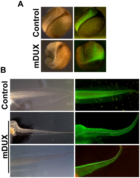
Figure 4. mDUX expression in Xenopus.
(A) Neurula stage embryos following injection of GFP mRNA alone (control) or GFP+mDUX mRNA. The green color indicates the domain filled by the RNA injection. The control has a normal neural plate while the mDUX case is very abnormal due to deranged gastrulation movements. (B) Tail muscle pattern in stage 45 tadpoles. The green color shows immunostaining with 12/101 antibody. Control shows the normal pattern of myotomes. Two examples (mDUX) demonstrate how injection into blastomeres V2.1 and 2.2 results in an inhibition of muscle differentiation on the injected (left) side.

