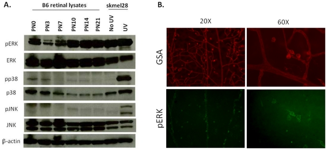
Figure 2. MAPK is active and spatially expressed in developing retinal vasculature.
(A) Western blot analysis of MAPK pathway activity during retinal vascular development. Eyes were collected and retinal lysates were prepared from C57BL/6 mice at the postnatal (PN) days indicated. Lysates from untreated (No UV) or UV-treated (UV) SK-MEL-28 cells were used as a positive control for MAPK activation. Immunoblotting shows strong expression of phosphorylated ERK (pERK), especially during times of angiogenic vascular growth in the retina, while phosphorylated p38 (pp38) and JNK (pJNK) are weakly expressed at earlier timepoints. (B) Epifluorescent images at low (20×) and high (60×) magnification of PN10 retinas stained with GSA (red) and pERK (green), to indicate pERK staining specific to the sprouting vessels.

