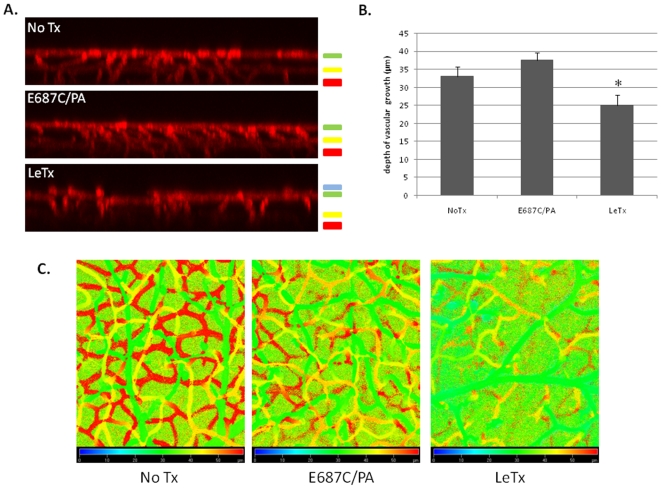
Figure 4. LeTx treatment delays growth of retinal vasculature into the inner plexus.
Mice at PN10 were intravitreally injected either with no treatment (No Tx), E687C/PA (0.02 µg E687C and 0.1 µg PA in 1 µL) or LeTx (0.02 µg LF and 0.1 µg PA in 1 µL). Eyes were collected 4 days post treatment, stained with GSA and flat mounted for confocal microscopy. (A) z-axis projection of GSA stained retinas show loss of inner plexus growth following LeTx treatment. The three layers of radial vascular growth are indicated by color bars to the right of the images, which correspond to the depth coding images in (C). While the superficial layer (green; a depth of approximately 30 µm from the top of the stack) is maintained in all treatments, there is a lack of intermediate plexus (yellow; a depth of 45 µm) and deep plexus (red; a depth of 55–60 µm) growth in the LeTx treated retina (A and C). (B) Depth of vascular growth from the superficial layer (green in A and C) through the deep plexus (red in A and C) was quantified from analysis of z-stack sections. Error bars indicate standard deviation from a minimum of three separate experiments. * p<0.05 compared to E687C/PA treatment.

