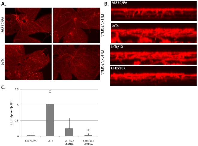
Figure 8. LeTx-induced neovascular tuft formation is VEGF-dependent.
Mice at PN10 were intravitreally injected either with E687C/PA (0.02 µg E687C and 0.1 µg PA in 1 µL), LeTx (0.02 µg LF and 0.1 µg PA in 1 µL), or LeTx in conjunction with 1× VEGF-neutralizing antibody (LeTx/1X; 15 pg/mL VEGFNA) or 10× VEGFNA (LeTx/10X; 150 pg/mL). Eyes were collected 8 days post treatment, stained with GSA and flat mounted for microscopic analysis. (A) Epifluorescent images of retinal wholemounts of either E687C/PA treated, LeTx treated, or LeTx plus VEGFNA treated retinas (LeTx/1X VEGFNA and LeTx/10X VEGFNA). (B) z-axis projection of GSA stained retinas show loss of neovascular growth following increasing addition of VEGF-neutralizing antibody to LeTx treatment. (C) The number of neovascular tufts in retinal whole mounts for each treatment was quantified using Imagine_0.16 software and plotted as # tufts/pixel2. Error bars indicate standard deviation from a minimum of three separate experiments. * p<0.02 compared to E687C/PA treatment. # p<0.03 compared to LeTx treatment.

