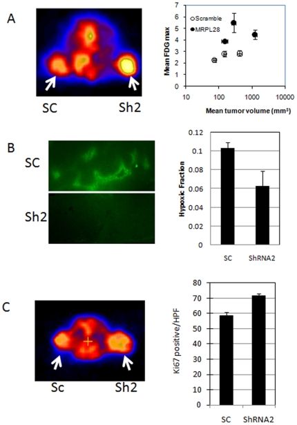
Figure 5. Metabolic changes cause accelerated growth the MRPL28 knockdown tumors.
Figure 5A Glucose uptake by microPET determination of 18F labeled deoxyglucose. Animals bearing one control scrambled ShRNA tumor and one MRPL28 knockdown SU86 tumor were injected with 200 uCi FDG; and imaged one hour later. Left panel shows heat map with MRPL28 tumors with increased uptake. Right panel shows quantitation of FDG uptake is increased independent of tumor size. Figure 5B) Size matched control Scrambled ShRNA SU86 and MRPL28 knockdown tumor bearing animals were injected with pimonidozole and 3 hours later sacrificed. Tumors were sectioned and stained for pimonidozole binding. Representative sections are shown in left panel and quantitation of hypoxic fraction shown in right panel. Figure 5C) Tumor proliferation in vivo was determined by 18F labeled thymidine for cellular proliferation. Animals bearing one control scrambled ShRNA and one MRPL28 knockdown SU86 tumor were injected with 200 uCi of 18F-thymidine and two hours later imaged. The PET signal was marginally increased, so tumor cell proliferation was confirmed by Ki67 staining of sections taken from the tumors. Right panel reports Ki67 positive cells per high powered field.

