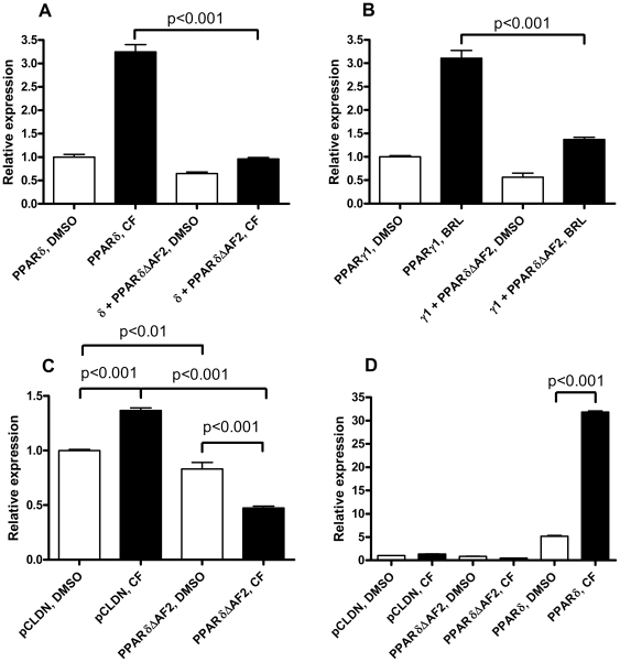
Figure 2. PPARδΔAF2 represses (A) PPARδ and (B) PPARγ1 signalling. (C) PPARδΔAF2 represses TK-promoter activity in a ligand-enhanced fashion.
COS-1 cells were transiently transfected with (per well in six-well plates) (A) 50 ng pCLDN-hPPARδ or (B) pCDLN-hPPARγ1 and 250 ng pCLDN or pCLDN-hPPARδΔAF2. (C) and (D) T47D cells were transfected with (per well in a six-well plate) 500 ng pCLDN, pCLDN-hPPARδΔAF2 or pCLDN-hPPARδ. (D) is identical to (C) except for the two additional bars representing over-expression of PPARδ with and without CF.

