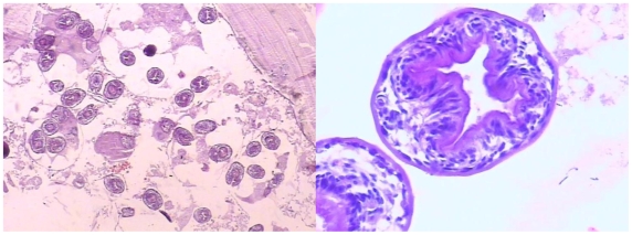
Figure 2. Histopathologic section (haematoxylin and eosin stain) of the liver lesion from a ground squirrel (Spermophilus dauricus/alashanicus).
Left panel shows the typical appearance of a fertile Echinococcus granulosus cyst with laminated and germinal layers, brood capsules and numerous viable protoscoleces; right panel is an enlarged part of the section showing two viable protoscoleces (×1000).

