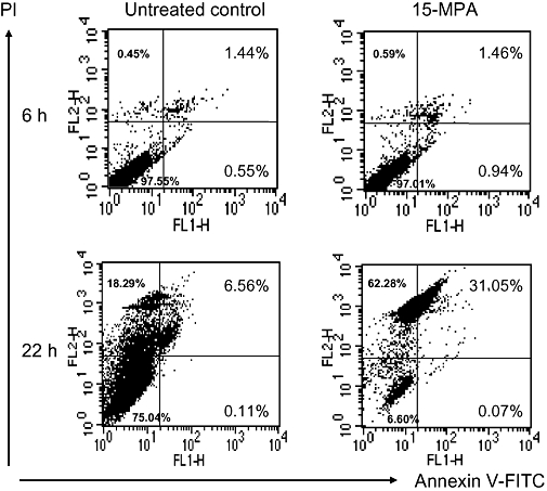
Figure 5.

Flow cytometric analysis of BV2 cells treated with 15-methoxypinusolidic acid (15-MPA). BV2 cells were treated with 15-MPA (50 µmol·L−1) for 6 or 22 h. Apoptotic cells were detected by staining cells with propidium iodide and anti-annexin V antibody-conjugated with fluorescein isothiocyanate (FITC) followed by flow cytometry. The percentage of the number of cells for each quadrant is shown (mean ± SD of data from three independent experiments).
