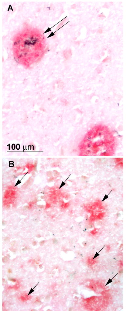
Figure 1.
Photomicrographs of neuritic (A, double arrow) and diffuse plaques (B, single arrows), from adjacent areas of the temporal cortex of a case that was rated npAD with the Khachaturian and Washington University criteria, probable AD with CERAD criteria, and intermediate probability of AD with NIA-Reagan criteria. Stained with a double immunohistochemical procedure in which Aβ is stained red and paired helical filaments are stained black.

