
Figure 6. Overexpression of PTP1B stabilizes VE-cadherin-mediated cell-cell adhesion and reduces VE-cadherin tyrosine phosphorylation.
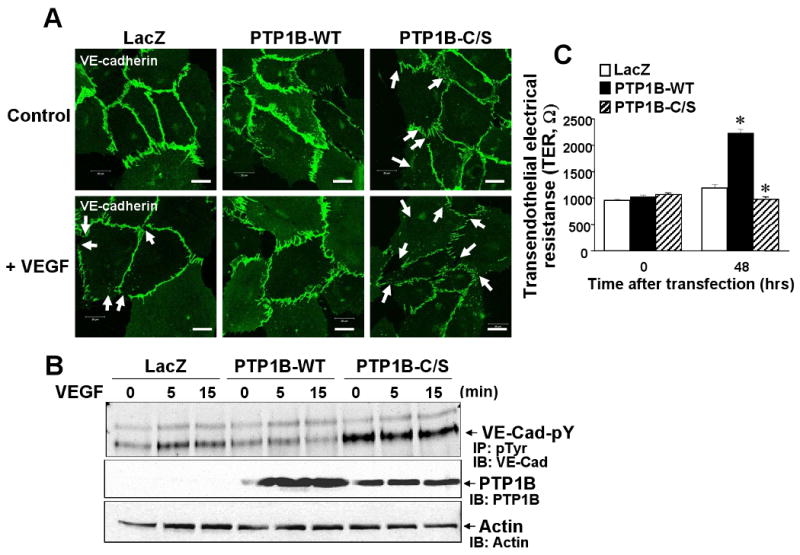
HUVECs infected with Ad.LacZ, PTP1B-WT or PTP1B-C/S were stimulated with VEGF (50 ng/mL). A, Cells were permeabilized, fixed and stained with anti-VE-cadherin antibody, followed by FITC-conjugated goat anti-rabbit antibody. White arrows show VEGF-induced decrease in VE-cadherin staining at cell-cell contact with formation of small gaps. Images were taken at 5 different fields/well, and representative of more than 3 independent experiments. B, Lysates were immunoprecipitated with anti-pTyr antibody, followed by immunoblotting with VE-cadherin antibody (Upper). Lysates without immunoprecipitation were immunoblotted with anti-PTP1B antibody (bottom). Results are representative of 3 independent experiments. C, Real-time measurement of transendothelial electrical resistance (TER). Values are the mean ± SE for 3 independent duplicate experiments. *P < 0.05 vs. Ad.LacZ.