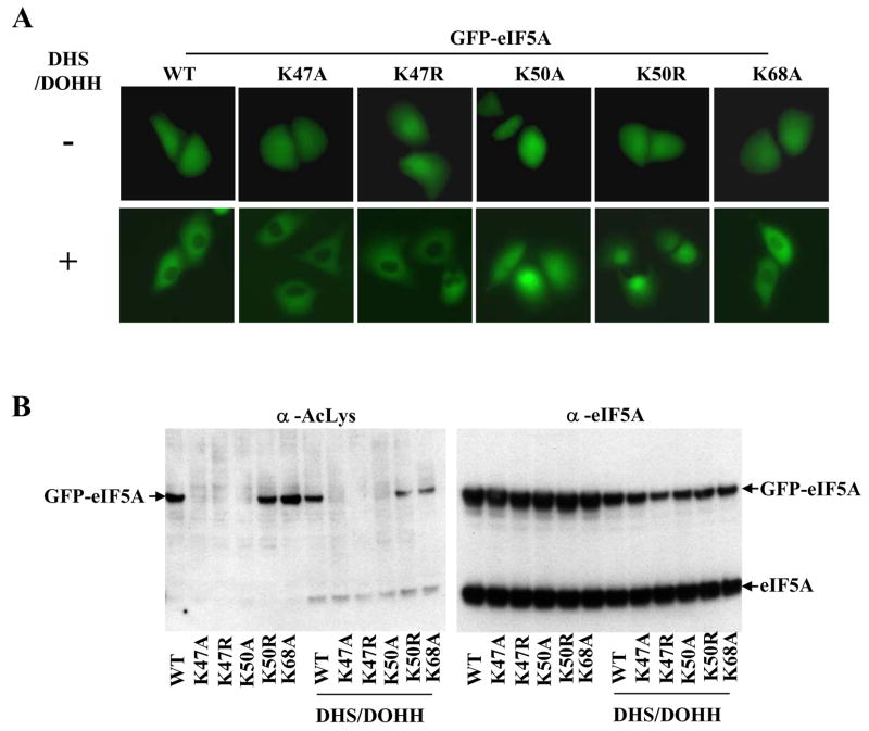
Fig. 4. Comparison of the cellular distribution of GFP-eIF5A mutant proteins.
(A) direct visualization of GFP fluorescence after 48h of transfection. HeLa cells were transfected with vectors encoding GFP-eIF5A wt and mutants, without (upper panels) or with (lower panels) vectors encoding DHS and DOHH. (B) western blots of the above samples using AcLys and eIF5A antibodies (BD Bioscience).

