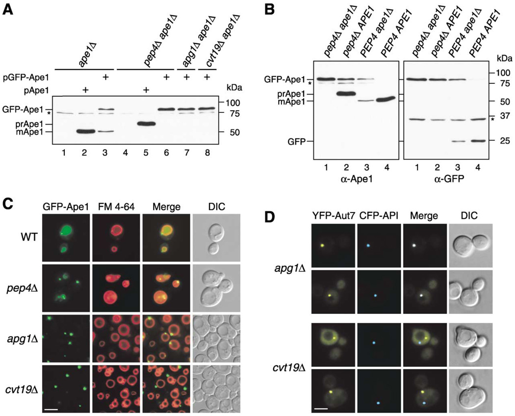
Figure 1. GFP-Ape1 Travels to the Vacuole via the Cvt Pathway.
(A) Cell extracts from ape1 A (lanes 1–3), pep4Δ ape1Δ (lanes 4–6), apg1Δ ape1Δ (lane 7), and cvt19Δ ape1Δ (lane 8) strains containing either empty vector (pRS414; lanes 1 and 4), pApe1 (lanes 2 and 5), or pGFP-Ape1 (lanes 3, 6, 7, and 8) were resolved by SDS-PAGE. Western blots were performed with anti-Ape1 antiserum.
(B) Western blot of cell extracts from pep4Δ ape1Δ (lane 1), pep4Δ (lane 2), ape1Δ (lane 3), and wild-type (lane 4) strains containing pGFP-Ape1 with anti-Ape1 (left panel) and anti-GFP (right panel) antisera. An asterisk (*) indicates nonspecific bands.
(C) Localization of Ape1 by fluorescence microscopy of wild-type, pep4Δ, apg1Δ, and cvt19Δ strains expressing GFP-Ape1. Cells were grown to midlog phase and labeled with FM 4-64 in SCD medium. Bar, 5 µm.
(D) CFP-Ape1 colocalizes with YFP-Aut7 in apg1Δ cells, but not in cvt19Δ cells. The apg1Δ and cvt19Δ cells coexpressing CFP-Ape1 and YFP-Aut7 were grown in SCD medium to midlog phase and examined with fluorescence and DIC microscopy. Bar, 5 µm.

